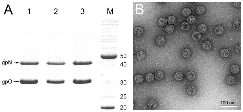
Figure 4.
Expression of mutant gpO. (A) Coomassie-stained SDS-PAGE showing expression of gpN and gpO from the clones O(D19A)+N (lane 1), O(H48A)+N (lane 2) and O(S107A)+N (lane 3). Lane M, marker; MW as indicated (kDa). (B) Electron micrograph of negatively stained procapsid particles produced in the O(D19A)+N co-expression. Scale bar, 100 nm.

