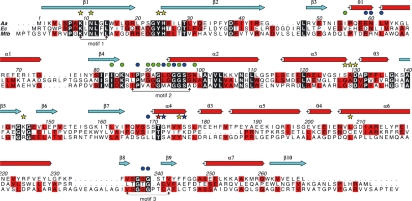
Fig. 7.
Primary and secondary structure of AaIspE and sequence alignment of A. aeolicus (Aa), E. coli (Ec) and M. tuberculosis (Mtb) orthologues. Residues conserved in all three sequences are boxed in black, those conserved in two of the three sequences are boxed in red. Three GHMP kinase superfamily conserved motifs are marked. The secondary structure of AaIspE is shown with helices as red cylinders and strands as cyan arrows. Residues marked with a star interact with substrate (yellow indicates a direct interaction, blue interactions bridged by water molecules). Thr171 is marked with a red star to indicate that it interacts with both CDPME and ADP. Residues marked with a dot interact with ADP (green indicates a direct interaction, blue water mediated interactions).

