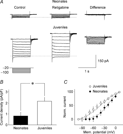
Figure 5. The Kv7/M channel enhancer retigabine potentiates.
Im more in juvenile than in neonatal neurons Examples of deactivating currents recorded from a P1 (upper traces, Neonates) and from a P18 (lower traces, Juveniles) CA3 principal cell. The deactivation protocol (represented below the current traces) consisted in stepping the voltage from −20 mV down to −100 mV in increments of 10 mV, before (Control) and during bath application of 10 μm retigabine (Retigabine). On the right, retigabine-sensitive currents obtained by subtracting the currents recorded in the presence of retigabine from control (Difference). The horizontal dotted lines mark the Im reversal potential. B, each column represents the density (pA pF−1) of retigabine-sensitive currents (for voltage steps from −20 to −100 mV) normalized to the cell capacitances in neonatal (n = 5) and in juvenile neurons (n = 5). *P < 0.05. C, voltage dependence of deactivated retigabine-sensitive currents (normalized to the maximal currents obtained at +10 mV) obtained in neonatal (filled symbols) and juvenile neurons (open symbols). Continuous lines represent the Boltzmann fits of experimental data (each point is the average of 4–5 individual values). The V1/2 values were −16.6 ± 6.0 mV and −39.8 ± 4 mV for neonatal and juvenile neurons, respectively. These values were significantly different (P < 0.05). The corresponding slope factors were 21.4 ± 6.4 and 24.5 ± 3.2.

