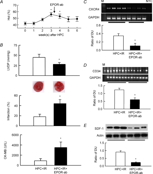
Figure 7. EPO receptor antibody (EPOR-ab) abolishes HPC-mediated functional protection in IR hearts.
A, EPOR-ab given 3 weeks (indicated by arrow) after HPC reduced the haematocrit. *P < 0.05 compared to week 0, i.e. before HPC induction. B, changes in LVDP, infarction with representative TTC-stained cardiac sections, and plasma CK-MB in untreated HPC + IR hearts or in hearts treated with EPOR-ab (n = 7 for each). *P < 0.05 compared to the HPC + IR group. C–E, upper representative blots showing CXCR4 mRNA, CD34 mRNA and SDF-1 protein expressions in 4, 7 and 3 hearts of each group, respectively. Lower bar graph shows the ratio of the DUs for CXCR4 and CD34 to GAPDH (n = 7 for each) and SDF-1 to actin (n = 6). M, 100 bp of DNA ladder. NTC, no template control. *P < 0.05 compared to the HPC + IR group.

