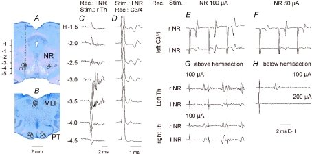
Figure 2. Location of the stimulating electrodes and descending rubrospinal volleys.
A and B, reconstruction of the stimulation sites in the red nuclei (NR) and in the pyramidal tracts (PT) in five experiments in which effects of NR stimulation were tested on motoneurones. They are displayed on representative brainstem sections in the plane of the inserted electrodes. Stimulation sites in the medial longitudinal fascicle (MLF) are projected on the same sections of the brainstem as the PT stimulation sites although they were a few millimetres more caudally. Stimulation sites to the left show those ipsilateral to the left side motoneurones recorded from in preparations with either ipsilateral or contralateral descending tracts intact. Stimulation sites to the right are contralateral with respect to the same motoneurones. The circles indicate location of the electrolytic marking lesions. The filled circle is for the data in C and D. The scale to the left of A shows Horsley-Clarke's horizontal coordinates. C, antidromic field potentials recorded along the electrode track indicated in A; they were evoked by 500 μA applied to the contralateral lateral funiculi. D, descending volleys recorded from the dorsal columns at the third cervical (C3) level when the stimuli (100 μA) were applied at the indicated depths. Note that the first one was evoked from locations within which antidromic field potentials were recorded (H-3 to -4) and from just outside the dorsal and ventral borders of the NR (H-2.5 and -4.5). The second one would correspond to synaptic activation of rubrospinal neurones but also of other neurones at unknown locations and/or destination activated from H-1 to -2.5. E and F, comparison of descending volleys evoked by 100 and 50 μA triple stimuli applied in the right and left NR at the locations at which maximal antidromic field potentials were evoked by Th stimuli as in C; these volleys were recorded from the border between the dorsal columns and the left lateral funiculus at a C3/4 level. Note that both were evoked at similar latencies as indirect volleys in D and that both were temporally facilitated. G and H, comparison of descending volleys evoked from the left and right NR above and below hemisection of the spinal cord made on the right side; they were recorded in parallel with descending volleys illustrated in E and F. Note that above the hemisection the volleys were evoked from both the left and right NR (being larger contralaterally) while below the hemisection they were evoked only from the right (contralateral) NR. Note also that they were evoked in a preparation in which only indirect volleys were evoked from either ipasilateral or contralateral NR and that both their latency and their temporal facilitation characterize them as evoked indirectly. All of the records are with the negativity up and with shock artefacts truncated.

