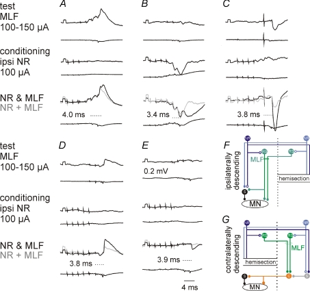
Figure 9. Ipsilateral NR neurones enhance ipsilateral actions evoked from the MLF via ipsilaterally and contralaterally descending pathways.
Upper traces, intracellular records from four motoneurones in 2 cats: DP (A), GS (B and D), PBST (C) and Sart (E). Lower traces, records from the cord dorsum in the L6 or L7 segment. In each column the records show effects of stimulation of the ipsilateral (A, B and C) or contralateral (D and E) MLF alone, of the ipsilateral NR alone and of the NR and the MLF together, with the sum of effects evoked by separate stimuli in grey. The latencies of the facilitated components shown in bottom panels are from the last MLF stimulus. The records are from preparations with only the ipsilaterally (A–C) or only the contralaterally (D and E) descending pathways left intact, with the corresponding simplified diagrams of the neuronal networks in F and G.

