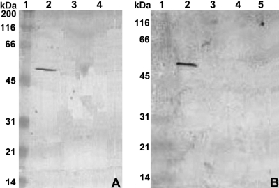
FIG. 3.
Western blot analysis of LsdB secretion by G. diazotrophicus. Cells were grown with 0.1% fructose plus 1% glycerol, except in blot A, lane 4. Supernatant proteins separated by SDS-PAGE were probed with anti-LsdB polyclonal antibody. Blot A lanes: 2, wild-type SRT4; 3, lsdA::nptII-ble mutant AD1; 4, SRT4 with 0.5% glucose replacing glycerol (negative control). Blot B lanes: 2, SRT4; 3, gspG::nptII-ble mutant AD6; 4, gspO::nptII-ble mutant AD8; 5, gspF::nptII mutant AD9. Lane 1 in both blots corresponds to the prestained broad-range protein marker from New England Biolabs.

