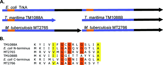
FIG. 1.
Schematic of aligned TrkA proteins. (A) The schematic shows the nature of the two-subunit TrkAs of T. maritima and M. tuberculosis CDC1551 in relation to the E. coli TrkA. The blue regions are the KTN/RCK domains. The characteristic nucleotide binding motifs GXGXXG are shown in orange. (B) An alignment of the nucleotide binding regions. Orange residues are the conserved glycines involved in nucleotide binding. The yellow residues are the extended conserved residues which indicate NAD(H) binding as described previously (20). TM1088B does not have the complete binding motif.

