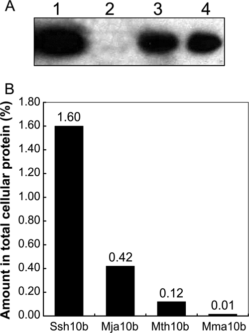
FIG. 1.
Expression levels of Sac10b homologs determined by Western blotting. (A) Western blotting of Mma10b. Proteins were separated with a 12% SDS-polyacrylamide gel and then transferred onto a PVDF membrane. The membrane was incubated sequentially with rabbit anti-Mma10b antiserum and anti-rabbit IgG-horseradish peroxidase conjugate. The proteins were detected with enhanced chemiluminescent substrates and exposed to CL-XPosure film for 5 s. Lane 1, 2, and 3, loaded with 400 μg of protein crude extracts of S2 (wild type), S590 (the Δmma10b mutant), and S591 (the complemented Δmma10b mutant), respectively. Lane 4, loaded with 25 ng of recombinant Mma10b. (B) Amounts of Sac10b homologs in wild-type cells as determined by Western blotting. Percentages of Sac10b homologs in total cellular protein were the averages of two independent measurements.

