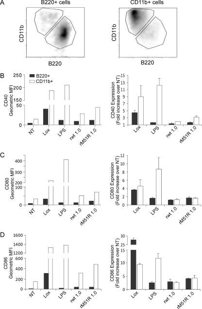
FIG. 3.
Separated B220+ and CD11b+ populations in F-DC respond to rwt and rM51R-M viruses. The B220+ and CD11b+ populations in F-DC were separated and infected with rwt or rM51R-M virus at an MOI of 1 PFU/cell or treated with Lox or LPS for 24 h. After infection, the expression of costimulatory molecules was measured in combination with the subset markers CD11b or B220 by flow cytometry. The efficiency of separation of the CD11b+ and B220+ DC populations is shown in panel A. The cell surface expression of CD40 (B), CD80 (C), and CD86 (D) in each sample is shown as the geometric mean fluorescence (left side) and is representative of two individual experiments. The increase in costimulatory molecule expression over that in untreated cells (NT) was quantitated (right side). The data are the means ± the standard deviations for two experiments.

