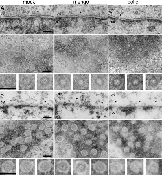
FIG. 2.
Electron microscopy of nuclear pores in infected cells. HeLa-3E cells were infected with mengovirus and poliovirus and fixed at 4 h p.i. The upper rows in panels A and B correspond to cross-sectioned nuclear envelopes, the middle rows are tangentially cut nuclear envelopes in plane, and the lower rows represent the twofold-enlarged examples of NPC from the appropriate middle rows. Scales bars correspond to 200 nm. The nuclear sides in cross-sections are marked as “nuc,” and the cytoplasmic ones are marked as “cyt.” Regions of NPC are marked with asterisks. (A) In situ fixation. The bar-like structures visible in NPC cross-sections of healthy cells are absent from a number of pores after poliovirus and mengovirus infections. In tangential section, control NPC display an electron-dense granule in the central channel, which is totally or partially lost in infected cells. (B) Fixation after dissolution of membranes with Triton X-100. NPC attached to the lamina are visible in cross-sections of the control envelope, and these complexes appeared to be only slightly disorganized in the mengovirus-infected sample. On the other hand, they are significantly destroyed by poliovirus infection. In tangential sections, the pores in control and mengovirus-infected cells looked very similar, whereas the NPC from poliovirus-infected cells displayed a much higher electron transparency and are less structured.

