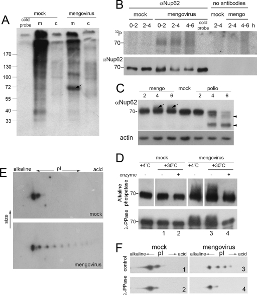
FIG. 3.
Nup62 is hyperphosphorylated in mengovirus-infected cells. (A) [32P]phosphate incorporation in WGA-binding proteins upon cardiovirus infection. HeLa-3E cells were labeled with [32P]orthophosphate during the first 2 h of infection, lysed, and subfractionated into cytosolic (c) and membrane (m) fractions as described in Materials and Methods. Nups were precipitated with WGA-beads and analyzed by PAGE and autoradiography. The first lane represents the control probe incubated for 2 h in ice. The arrow points out to a ∼70-kDa band appearing in mengovirus-infected cells. (B) Immunoprecipitation of 32P-labeled Nup62 from mengovirus-infected cells. Infected and mock-infected HeLa-3E cells were labeled with inorganic [32P]phosphate during the time periods indicated. The cold probe was exposed to the label for 6 h in ice. The cells were lysed and Nup62 was immunoprecipitated and subjected to electrophoresis as described in Materials and Methods (upper row). The amounts of Nup62 in the samples were controlled by Western blotting (lower row). (C) Modifications of Nup62 by different picornaviruses. HeLa-3E cells were infected with mengovirus or poliovirus for indicated times, and their extracts were investigated by PAGE, followed by Western blotting with anti-Nup62 antibodies. The lower mobility component of Nup62 from mengovirus-infected cells are marked with arrows; higher-mobility bands from poliovirus-infected cells corresponding to degradation products are marked with triangles. (D) The low-mobility forms of Nup62 disappeared from mengovirus-infected cell after alkaline phosphatase (upper row) and λ-PPase (lower row) treatments. Lysates from mock- and mengovirus-infected cells made at 4 h p.i. in the case of the former enzyme and immunoprecipitated preparations of Nup62 from such lysates in the case of the latter were treated with the phosphatases for 30 min at 30°C and analyzed by Western blotting. (E) Detection of hyperphosphorylated forms of Nup62 in mengovirus-infected cells. Cell lysates were prepared as in panel C and were subjected to 2D electrophoresis followed by Western blotting. (F) The additional acidic spots of Nup62 disappeared after treatment with λ-PPase. Samples 1 to 4 from the lower row of panel D were subjected to 2D electrophoresis, followed by Western blotting.

