
FIG. 8.
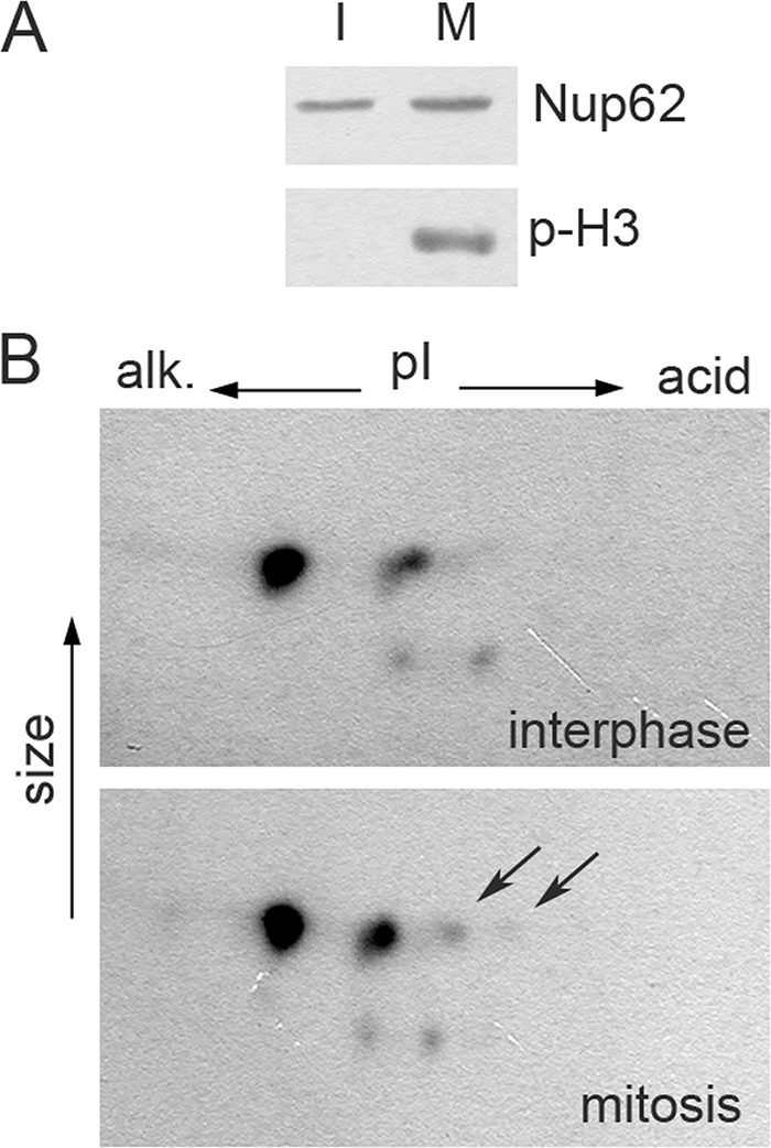
Nup62 is phosphorylated in mitotic HeLa cells. (A) Control for synchronization. Samples of interphase (I) and mitotic (M) cells (see Materials and Methods) were analyzed by Western blotting for Nup62 and Ser-10 phosphorylated H3 histone (p-H3). (B) The same preparations were analyzed by 2D electrophoresis, followed by Western blotting for Nup62. Acidic spots are visible in the preparations of mitotic cells, but those of interphase cells are marked by arrows.