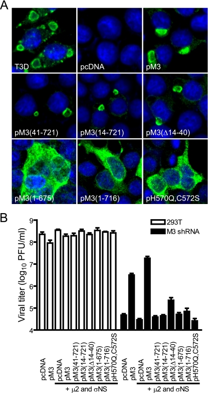
FIG. 1.
trans-complementation of reovirus replication in cells expressing M3 (μNS-specific) shRNA. (A) Subcellular localization of mutant μNS proteins. 293T cells were infected with T3D at an MOI of 2 PFU/cell or transfected with the indicated plasmid constructs, and μNS expression was examined 24 h later by confocal immunofluorescence microscopy using μNS-specific antiserum (green). Nuclei were stained with TO-PRO3 (blue). pcDNA, nonrecombinant vector control; pM3, encodes full-length μNS; pM3(41-721), encodes μNS amino acids 41 to 721; pM3(14-721), encodes μNS amino acids 14 to 721; pM3(Δ14-40), encodes μNS amino acids 1 to 13 and 41 to 721; pM3(1-675), encodes μNS amino acids 1 to 675; pM3(1-716), encodes μNS amino acids 1 to 716; pH570Q, H572S, encodes full-length μNS containing His-to-Gln and His-to-Ser amino acid substitutions at positions 570 and 572, respectively. (B) Viral growth in cells expressing vector-encoded μNS proteins. Parental or M3 shRNA-expressing 293T cells were transfected with plasmids expressing wt or mutant μNS proteins, wt μ2, or wt σNS as indicated, followed by infection with T3D at an MOI of 10 PFU/cell. At 24 h postinfection, viral titers were determined by plaque assay. Results are mean viral titers from three independent experiments. Error bars denote standard deviations.

