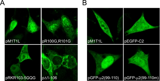
FIG. 6.
Requirement for conserved N-terminal basic residues in μ2 subcellular localization. (A) Intracellular distribution of wt and mutant μ2 proteins. 293T cells were transfected with plasmid constructs encoding the indicated T1L-derived μ2 proteins, which were detected by confocal immunofluorescence microscopy using μ2-specific antiserum (green). pM1T1L, wt T1L μ2; pR100G,R101G, μ2 containing Arg100-to-Gly and Arg101-to-Gly substitutions; pRKR103-5GQG, μ2 containing Arg103-to-Gly, Lys104-to-Gln, and Arg105-to-Gly substitutions; pΔ1-106, truncated μ2 protein lacking the N-terminal 106 amino acid residues. (B) Intracellular distribution of GFP fused to μ2 protein sequences. L cells were transfected with the indicated plasmid constructs. Expression of wt T1L μ2 protein was detected by confocal immunofluorescence microscopy using μ2-specific antiserum (green). Intrinsic GFP fluorescence was detected using confocal microscopy (green). pEGFP-C2, enhanced GFP; pGFP-μ2(99-110), EGFP appended at the C terminus with the μ2 putative nuclear localization sequence, D99RRLRKRLMLKK110; pGFP-μ2(99-110m), pGFP-μ2(99-110) containing Ala substitutions for μ2 basic residues.

