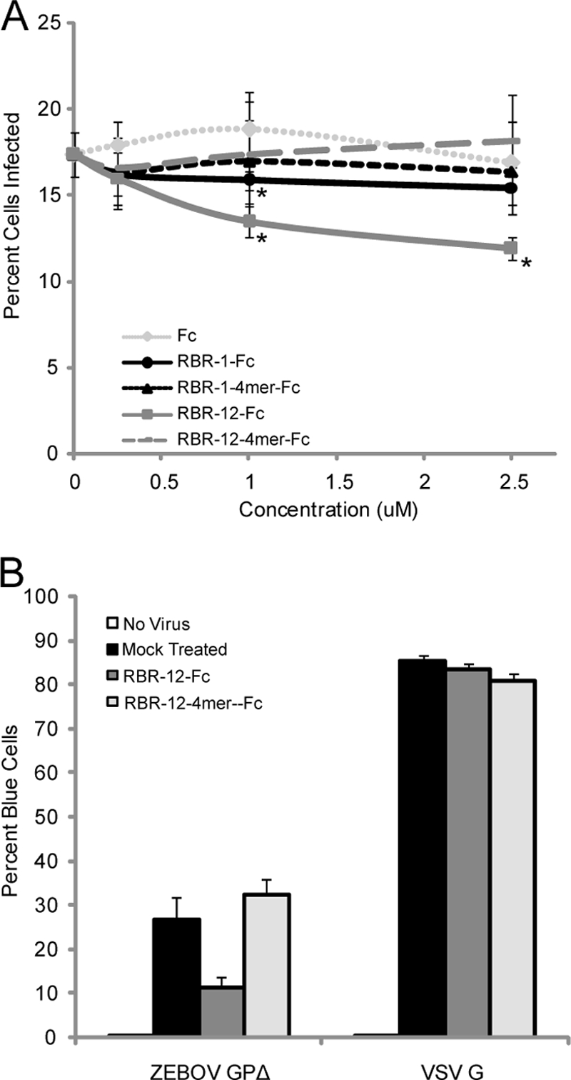
FIG. 8.

Inhibition of EBOV GP1,2Δ-mediated infection by RBR-1-Fc and RBR-12-Fc. (A) Vero E6 cells were incubated for 3 h with VSVgfp-GP1,2Δ in the presence or absence of RBR-1-Fc, RBR-12-Fc, or control rabbit Fc at the indicated concentration. Unbound virus was removed by washing, the cells were incubated overnight, and GFP expression was quantified by flow cytometry. Samples were analyzed in triplicate. The average values from one representative experiment are shown. Error bars represent standard deviations. Significance (relative to the rabbit Fc control tested at the same concentration) was determined by Student's t test. *, P < 0.05. The exact same experiment was conducted two times with similar results. RBR-1-Fc and RBR-12-Fc were tested five additional times (with HIV gp120-Fc as a negative control), with very similar results. (B) Vero E6 cells were infected with HIVblam-GP1,2Δ or HIVblam-G in the presence or absence of WT or 4mer mutant RBR-12-Fc at 800 nM. Cells were loaded with the beta-lactamase substrate CCF2/AM. Cells loaded only with CCF2/AM served as a negative control (no virus). The extent of CCF2/AM cleavage by beta-lactamase introduced into the cytoplasm was evaluated by flow cytometry (detected by the change in dye emission from green to blue). Samples were analyzed in duplicate. The averages for duplicate samples from one representative experiment are shown. Error bars represent standard deviations.
