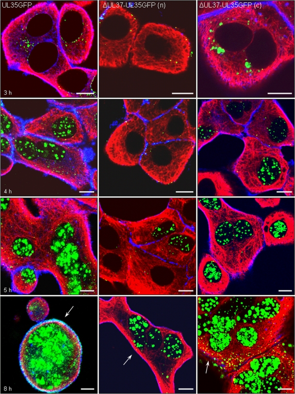
FIG. 6.
pUL35 expression, capsid formation, and viral egress. RK13 cells infected at an MOI of 200 with PrV-UL35GFP (left panels), noncomplemented PrV-ΔUL37/UL35GFP (n) (middle panels), or phenotypically complemented PrV-ΔUL37/UL35GFP (c) (right panels) were fixed 3 h (top row), 4 h (second row), 5 h (third row), and 8 h (bottom row) after infection. Direct and indirect fluorescence reactions were analyzed by confocal laser-scanning microscopy. Green, pUL35-eGFP; red, MT; blue, actin. Egressing viral capsids at the cell periphery are marked by arrows. Bar, 10 μm.

