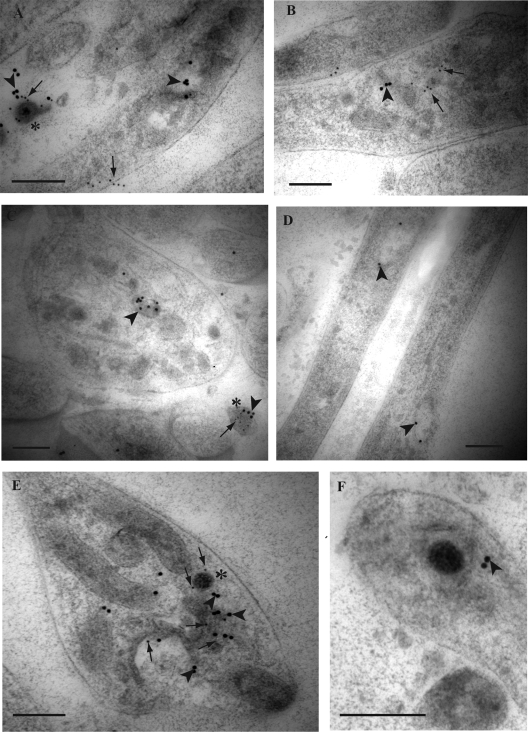
FIG. 2.
Double immunogold labeling for HSV-1 tegument protein VP16 and envelope gB in axons of HSV-1-infected human DRG. Coverslips with DRG cultures were processed to Lowicryl HM20, and dual immunogold labeling of ultrathin sections of mid and distal axons containing varicosities and growth cones was performed as described in Materials and Methods. Label for VP16 was detected with a 10-nm gold conjugate and for gB with a 15-nm gold conjugate. (A) Label for gB (15-nm gold particles; arrowheads) is associated with a large clear-cored vesicle in a varicosity. Label for VP16 (10-nm gold particles; arrows) is found on the plasma membrane separately from label for gB (arrowhead). Note the label for both gB (arrowhead) and VP16 (arrow) is found on the extracellular viral particle (indicated by the asterisk) adjacent to the varicosity. (B) Label for gB (arrowhead) and VP16 (arrows) on separate dense-cored vesicles in a varicosity. (C) Label for gB (arrowhead) in a large dense-cored vesicle in a growth cone. Note the extracellular viral particle (indicated by the asterisk) adjacent to the growth cone is decorated with label for both gB (arrowhead) and VP16 (arrow). (D) Label for gB (arrowheads) on two separate large tubulovesicles in mid axons. (E) Label for gB (arrowheads) and VP16 (arrows) is present on tubulovesicular membrane structures in close proximity to an unenveloped capsid (indicated by the asterisk) in a growth cone. Label for VP16 (arrows) is also present on the unenveloped capsid. (F) Unenveloped capsid partially surrounded by a tubulovesicular membrane structure with associated label for gB (arrowhead). Bars, 200 nm.

