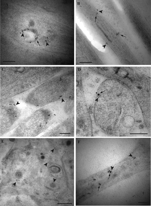
FIG. 3.
Immunogold labeling for TGN-46, GAP-43, Rab3A, tegument proteins VP16 and VP22, and envelope gB in mid and distal axons of HSV-1-infected human DRG. Ultrathin sections were simultaneously incubated with primary antibodies overnight. Detection of primary antibodies was obtained either by incubation with gold-conjugated antibodies of different sizes of gold particles (5 and 10 nm) or by incubation with secondary antibodies conjugated with 2-nm gold particles and sequential silver enhancement to produce two different sizes of gold particles. (A) Label for VP22 (arrowheads) and TGN-46 (arrows) together on tubulovesicular structures in a mid axon. (B) Label for gB (arrowheads) together with label for TGN-46 (arrows) on the plasma membrane in a mid axon. (C) Label for GAP-43 (arrowheads) is present on the plasma membrane of the tips of axons. (D) Label for GAP-43 (small gold particles) (arrowhead) is present in association with label for VP16 (large gold particles; arrow) on the plasma membrane of a growth cone. (E) Label for Rab3A (arrowheads) is found on clear and dense-cored vesicles as well as on the plasma membrane in a growth cone. Note that the label for Rab3A does not associate with the unenveloped capsid (indicated by the asterisk). (F) Label for VP16 (arrows) is present together with or separately from label for Rab3A (arrowheads) in an axon. Bars, 200 nm.

