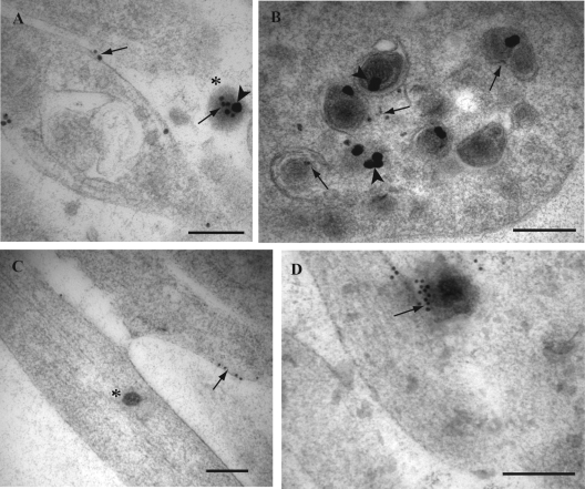
FIG. 5.
Immunogold labeling for GAP-43 and Rab3A in HSV-1-infected mid and distal human axons. Ultrathin sections were either single labeled for GAP-43 or Rab3A or dual labeled with monoclonal antibody to VP5 and rabbit polyclonal antibody for GAP-43 incubated simultaneously overnight. For double labeling, sequential incubation with secondary antibodies conjugated with 2-nm gold particles and silver enhancement was performed to produce two different sizes of gold particles. (A) Label for GAP-43 (small gold particles; arrows) is present on the plasma membrane of a varicosity and on the extracellular viral particle (indicated by the asterisk). The extracellular viral particle also carries label for VP5 (large gold particle; arrowhead). (B) Label for GAP-43 (arrows) is present on vesicles surrounding enveloped capsids in a growth cone while label for VP5 (arrowheads) decorates the enveloped capsids. (C) Label for GAP-43 along the plasma membrane of axons. Note that label for GAP-43 (arrow) does not associate with the unenveloped capsid (indicated by the asterisk) found in mid axon. (D) Label for Rab3A (10-nm gold particles; arrow) is present decorating an extracellular viral particle. Bars, 200 nm.

