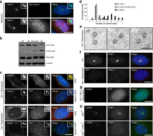
FIG. 6.
Cdk2 activity is required for assembly of centriole precursors. (a) CHO cells transiently transfected with myc-cyclin A were treated with mimosine for 48 h. Cells were stained for γ-tubulin (green) and myc (red). (b) Cell extracts were prepared from asynchronous (AS) and G0 (serum starved for 72 h) CHO cells or cells treated with mimosine or HU for 48 h. Western blots were probed with antibodies against pT14 Cdk2, pT160 Cdk2, total Cdk2, and anti-α-tubulin antibodies. (c) CHO and CHO:centrin1-GFP cells were treated with HU and the Cdk inhibitor olomoucine or roscovitine for 48 h. Cells were stained with γ-tubulin (red) and centrin or GFP (green) antibodies. (d) Histogram indicating the numbers of centrosomes detected by γ-tubulin staining in CHO cells treated for 18 h with HU (gray bars), for 18 h with HU followed by 30 h with HU and roscovitine (white bars), or for 48 h with HU (black bars). Standard deviations are indicated. (e) TEM analysis of CHO:centrin1-GFP cells treated with HU and roscovitine for 48 h reveals no duplication of centrioles and the absence of centriolar satellites. Bar, 500 nm. (f) CHO:centrin1-GFP cells were treated with HU or HU plus roscovitine for 48 h. Cells were stained with GFP (green) and PCM-1 (red) antibodies. (g) CHO:centrin1-GFP cells were treated for 48 h with HU, nocodazole, and roscovitine before being stained with γ-tubulin (red) and GFP (green) antibodies. (h) p53−/− and p53−/− Cdk2−/− MEFs were treated with HU for 48 h before being stained with γ-tubulin (red) and PCM-1 (green) antibodies. In panels a, c, f, g, and h, merge images include a DNA stain (blue). Bars, 10 μm.

