FIG. 3.
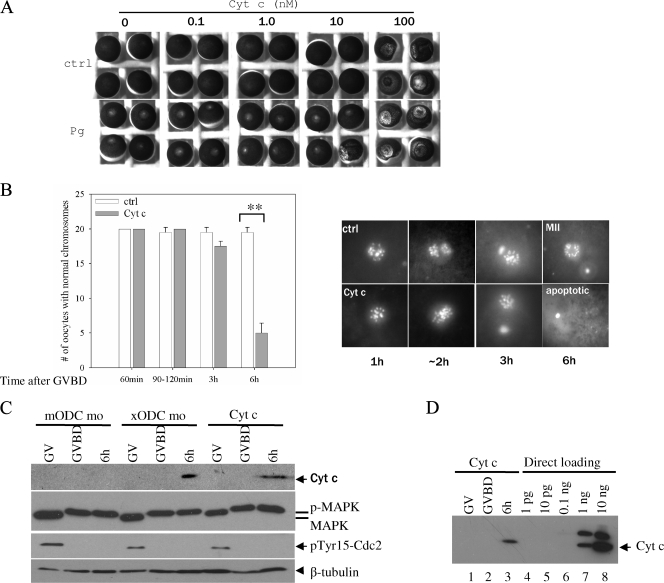
Cyt c injection caused apoptosis in metaphase II oocytes. (A) GV oocytes were injected with the indicated (final intracellular) concentrations of horse heart Cyt c. Half of the injected oocytes were incubated in OR2 (ctrl) and the other half in OR2 containing progesterone. Following overnight incubation, representative oocytes were photographed. Note the mottled phenotype in oocytes injected with 100 nM Cyt c, with or without progesterone treatment. (B) Control oocytes and oocytes injected with 0.1 nM Cyt c were treated with progesterone. GVBD oocytes were further incubated for the indicated periods of time before fixation for chromosome examination. **, P < 0.01 (Student's t test). (C) Oocytes injected with mODC mo (as a control), oocytes injected with xODC mo, and oocytes injected with 0.1 nM Cyt c were either untreated (GV) or treated with progesterone, withdrawn at GVBD or 6 h after GVBD. The oocytes were subjected to Cyt c release assays (top panel) or analyzed for MAP kinase phosphorylation (activation, as described for Fig. 2B) and for Cdc2 Tyr15 dephosphorylation (activation). The bottom panel depicts β-tubulin as a loading control. p-MAPK, phosphorylated (active) MAP kinase; MAPK, inactive MAP kinase. (D) The same three samples from the Cyt c-injected oocytes shown in panel C were reblotted (lanes 1 to 3, each representing five oocytes) together with the indicated amounts of horse heart Cyt c directly loaded on the gel (lanes 4 to 8). We estimate that about 500 pg of Cyt c (about one-half of the amount loaded in lane 7) was released from five oocytes (lane 3), assuming that the antibodies recognized Cyt c from the two species (94% identical in amino acid sequence) with equal levels of efficiency.

