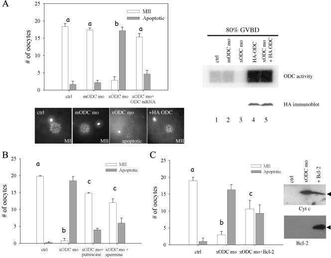
FIG. 4.
xODC mo-induced apoptosis can be rescued by HA-ODC, polyamines, and Bcl-2. (A) Control oocytes (ctrl) and oocytes injected with the indicated agents were treated overnight (15 to 20 h) with progesterone. Chromosome morphology was determined as indicative of normal metaphase II (MII) eggs or apoptosis. Shown are means (with SEM) for six experiments, with representative chromosome images below. Shown on the right are the results for an ODC activity assay (top) and an HA immunoblot analysis (bottom) of extracts, made when 80% of the oocytes of each group exhibited GVBD. Oocytes injected with HA-ODC mRNA alone were indistinguishable from control oocytes in terms of chromosome morphology (not shown). (B) Assays similar to those for panel A, except for inclusion, where indicated, of 5 mM putrescine or 5 mM spermine in OR2 medium at the same time as progesterone addition. Shown are means (with SEM) for three independent experiments. (C) Assays similar to those for panels A and B, with one group of oocytes injected with xODC mo and Bcl-2 mRNA. Shown are means (with SEM) for three independent experiments. On the right are representative immunoblots showing release of mitochondrial Cyt c (top) and expression of exogenous Bcl-2 (bottom). Differences in letters (a to c) denote significant differences in values (P < 0.01; Student-Newman-Keuls test).

