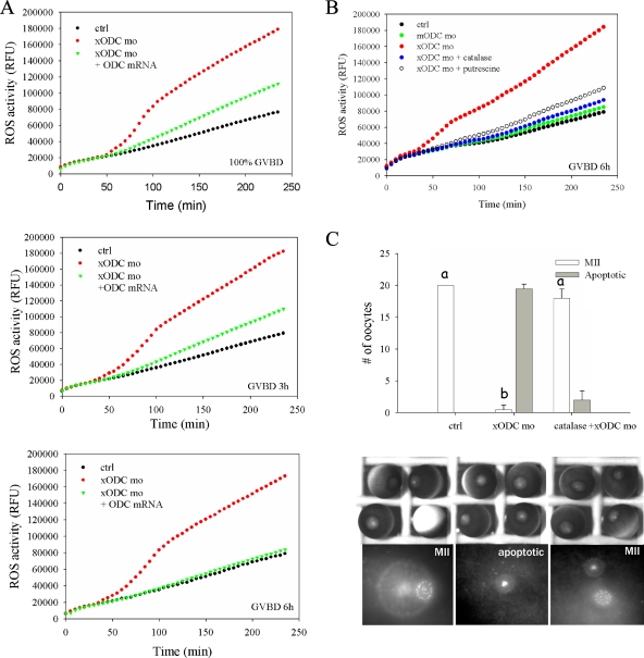
FIG. 5.
ODC deficiency resulted in elevated levels of ROS in oocytes. (A) Control oocytes (ctrl), oocytes injected with xODC mo, and oocytes injected with xODC mo plus HA-ODC mRNA were incubated in OR2 for 2 h before the addition of progesterone. Groups of 20 oocytes were withdrawn at the indicated times (at 100% GVDB or 3 h or 6 h after GVBD), lysed immediately, and assayed for ROS. The time points (in min) denote reaction times following the addition of carboxy-H2DCFDA. RFU, relative fluorescence units. (B) Uninjected oocytes (ctrl), oocytes injected with mODC mo, or oocytes injected with xODC mo alone or coinjected with catalase (350 ng per oocyte) were treated with progesterone. One group were injected with xODC mo and treated with progesterone in the presence of 5 mM putrescine. Oocytes in all groups were withdrawn 6 h after GVBD for ROS assays. (C) Control oocytes, oocytes injected with xODC mo, or oocytes coinjected with xODC mo and catalase (350 ng per oocyte) were incubated with progesterone overnight (15 to 20 h). Oocytes were fixed for chromosomal analyses. Shown are means (with SEM) for three independent experiments. Also shown are typical light images and chromosomal images of each group. One of the control oocytes was positioned upside-down, showing the vegetal hemisphere. Differences in letters (a and b) denote significant differences in values (P < 0.01; Student-Newman-Keuls test).

