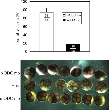
FIG. 6.
ODC deficiency caused embryo fragmentation. This graph shows percentages of normal embryos derived from xODC mo-injected or mODC mo-injected oocytes. Only cleaving embryos were included in the tally. The total numbers of embryos in the three experiments were also included in the graph. Shown below are representative images of embryos derived from host eggs (uncolored, middle row), embryos derived from xODC mo-injected oocytes (top row, exhibiting fragmentation), and those derived from mODC mo-injected oocytes (bottom row, exhibiting normal, albeit slightly slower, cleavage). All three groups of embryos were from the same fertilization. In other experiments, the two vital dyes were switched between the two groups of transferred eggs, with no significant difference in experimental outcome.

