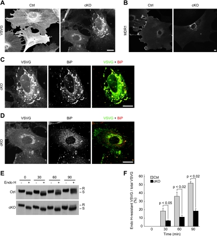
FIG. 10.
ER-to-Golgi complex protein transport is impaired in p31-deficient MEFs. (A, C, and D) p31flox/− MEFs were infected with Ad-LacZ (Ctrl) or Ad-Cre (cKO), followed by infection with another adenovirus encoding VSVG-GFP. Four days after the first adenovirus treatment, they were fixed 60 min after a temperature shift to a permissive temperature and double stained with anti-GFP and anti-BiP antibodies. The distribution of VSVG, which is detected by staining with the anti-GFP antibody, is shown in panel A. (C and D) Double-stained images of two cells in panel A (cKO) at higher magnification. (B) p31flox/+ (Ctrl) or p31flox/− (cKO) MEFs infected with Ad-Cre were infected with adenovirus encoding MDR1-GFP and were stained using anti-GFP antibody. (A to D) Bars, 10 μm. (E and F) VSVG-expressing control MEFs (Ctrl) and p31-deficient MEFs (cKO) were treated as described above, except that the cells were lysed, subjected to Endo H treatment, and analyzed by immunoblotting with a polyclonal anti-VSVG antibody. A representative experiment is shown in panel E. R and S denote Endo H-resistant and Endo H-sensitive forms of VSVG, respectively. (F) Ratio of Endo H-resistant VSVG to total VSVG (mean ± standard error of the mean) calculated from three independent samples. P values were determined by Student's t test.

