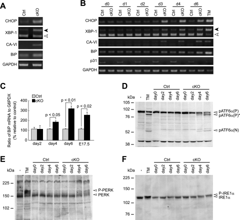
FIG. 4.
p31 deletion leads to ER stress. (A and B) RT-PCR analysis of ER stress-associated molecules in brains (A) and MEFs (B). Black and white arrowheads indicate spliced and unspliced forms of XBP-1, respectively. GAPDH was used as an internal control. (A) RT-PCR analysis of E17.5 brains from control mice (Ctrl) and CNS-specific knockout mice (cKO). (B) RT-PCR analysis of p31flox/+ (Ctrl) or p31flox/− (cKO) MEFs infected with Ad-Cre. “dX” represents samples prepared on day X after adenovirus treatment. As a positive control, MEFs were treated with tunicamycin for 24 h (TM). (C) Quantitative RT-PCR to evaluate level of BiP mRNA showed increased expression in p31-deficient MEFs (days 2, 4, and 6) and embryonic brains (E17.5). The ratio of BiP mRNA to G6PDX (glucose 6-phosphate dehydrogenase X-linked) calculated from three independent samples, were normalized against data of control. P values were determined by Student's t test. (D to F) Western blot analyses showed increased cleavage of ATF6α (D) and increased phosphorylation of PERK (E) and IREα (F). pATF6α(P), pATF6α(P)*, and pATF6α(N) indicate full-length ATF6α, nonglycosylated full-length ATF6α, and cleaved form of ATF6α, respectively.

