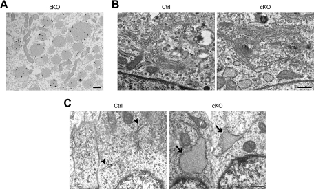
FIG. 8.
Characterization of abnormal ERs by electron microscopy. (A) Immunogold labeling of PDI was found in the vesiculated ER of p31-deficient MEFs. p31flox/− MEFs infected with Ad-Cre were observed 6 days after adenovirus treatment. (B) The Golgi complex is not disrupted in p31-deficient MEFs. p31flox/− MEFs infected with Ad-LacZ (Ctrl) or Ad-Cre (cKO) were observed 6 days after adenovirus treatment. (C) Enlargement of ERs (arrows) was also observed in neurons from the E16.5 brains of CNS-specific knockout mice (cKO) but not in neurons of control mice (Ctrl). Normal ERs are indicated by arrowheads. Bars, 500 nm.

