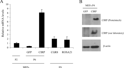
FIG. 2.
CIRP expression in various cell lines. (A) MEF cells were infected with a retroviral vector carrying the indicated genes, selected with appropriate antibiotics, and after selection were split following a 3T3 protocol. At P6 when clear phenotypic characteristics distinguish GFP- from CIRP-expressing cells, RNA was extracted to perform quantitative real-time PCR. Primary MEFs at early passage (P2) were also included, and ES cell lines CGR8 and ROSA 21 are shown as positive controls of CIRP expression as the original cDNA library was from both ES cell lines. (B) Duplicate dishes from CIRP- and GFP-expressing MEFs were kept for protein extraction. An immunoblot with anti-CIRP antibody is shown with two independent CIRP antibodies (one from Proteintech and the other generated in our laboratory). β-Actin is shown as loading control.

