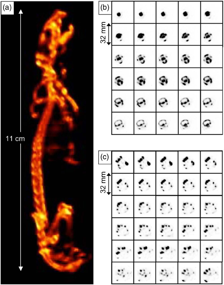
FIG. 7.
Volume rendering (a) from the SPECT images of a whole mouse skeleton and consecutive transaxial SPECT images of (bB) the mouse cranium from incisive bone to occipital squama, and (c) the mouse pelvic bone from ilium to ischiac bone with 0.5 mm separation between slices. 99mTc-MDP was used as the bone imaging agent.

