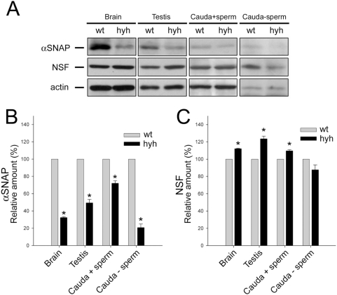
Figure 4. αSNAP and NSF expression in the reproductive tract of wild type (wt) and SP hyh (hyh) mice.
(A) Proteins extracted from testis and cauda epididymidis of wt and SP hyh mice were analyzed by Western blot using an antibody recognizing αSNAP (upper panel) or NSF (middle panel). Signals detected with an anti-actin antibody served as internal controls for equal protein loading (lower panel). Cauda epididymidis extracts were obtained before (Cauda+sperm) and after (Cauda-sperm) sperm were washed out from the organ. Brain was used as a positive control. Blots are representative of 3 or 4 independent experiments. (B, C) Densitometric analysis of Western blot for αSNAP (B) and NSF (C). Black bars (mean±SEM, N = 3 or 4) refer to the relative amount of each protein in hyh samples compared to wt. * p<0.05 (Student's t-test).

