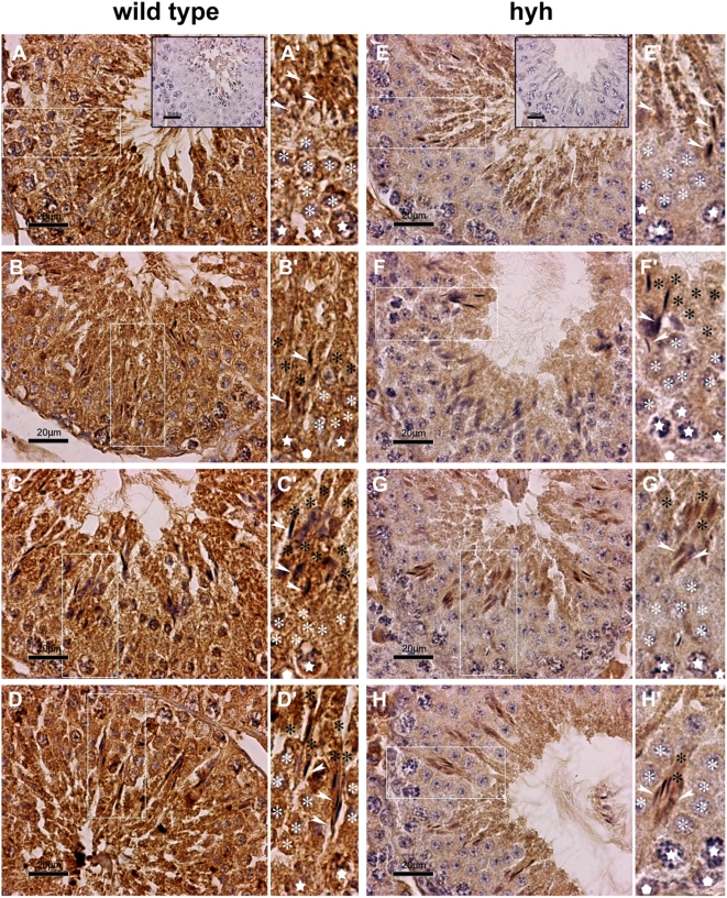
Figure 5. Immunolocalization of αSNAP in seminiferous epithelium.
Light micrographs of testis sections from wild type (A–D) and SP hyh mice (E–H) showing general and specific patterns of immunoperoxidase staining using an αSNAP-specific antibody. Background staining with hematoxylin. Different stages of epithelial maturation cycle in normal and mutant testis are presented in comparative mode. A'–H': High magnification images of the regions boxed in the corresponding panel (A–H). Epithelial polarization is oriented upwards. The symbols used are: spermatogonium (white pentagon); primary spermatocytes (white stars); round spermatids (white asterisk); elongated spermatids with high polarization, indicating the residual body or axonemal region (black asterisk) and the heads (white arrowheads). Controls without primary antibodies are shown in the inserts (A and E). In wild type animals, a strong immunoreaction was observed in the whole ephitelium. In contrast, mutant mice showed a notably diminished immunoreaction compared to that of wild type mice (compare E–H to A–D panels). This difference was not evident in spermatids undergoing elongation process. Scale bar, 20 µm.

