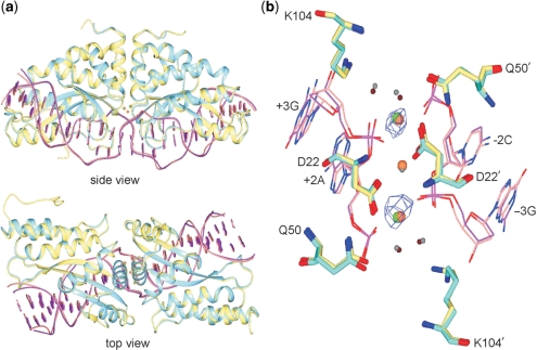
Figure 5.
Superposition of the I-MsoI:DNA and newly determined mMsoI:DNA co-crystal structures. (a) side and top views of the native homodimeric I-MsoI and newly determined monomeric mCreI co-crystal structures reveals backbone superposition with a 0.47 Å RMSD for protein and DNA. I-MsoI and mMsoI structures, and their DNA molecules, are shown respectively in cyan, yellow, purple and pink. Three calcium ions in I-MsoI:DNA structure and two calcium ions in mMsoI:DNA structure are shown, respectively, in green and coral. (b) Superposition of the I-MsoI and mMsoI active sites. Three active site residues—D22, Q50 and K104—with two calcium ions and two nucleotides flanking the scissile phosphodiester bond are shown. Four water molecules from I-MsoI:DNA and mMsoI:DNA structures are shown as small spheres in grey or tan, respectively. Anomalous difference mapping analysis revealed two calcium ions in the mMsoI:DNA active site. Figures were prepared using the CCP4 Molecular Graphics software package (33).

