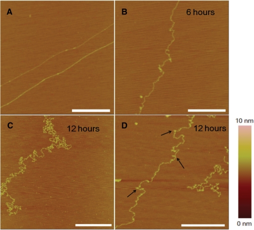
Figure 3.
AFM images of λ DNA alone or incubated with 77 μM cisplatin at different times. (A) Unmodified DNA in the absence of cisplatin. The DNA was stretched on mica surface (see Materials and methods section). (B) In the presence of cisplatin, 6 h incubation. (C) and (D) 12 h incubation. DNA coiling became more serious, accompanied by formation of tiny structures (micro-loops, as indicated by arrows). All scale bars are 500 nm.

