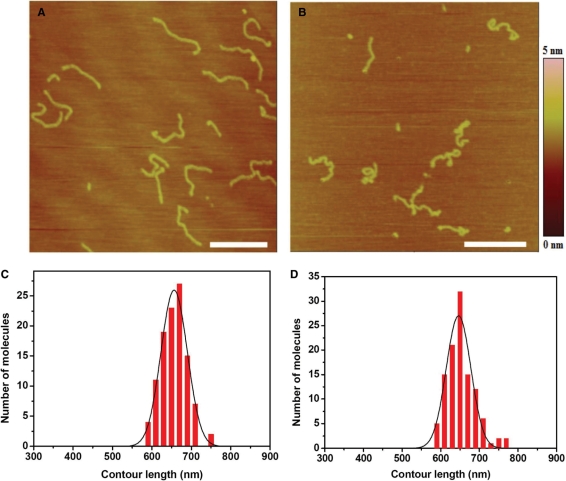
Figure 4.
(A) AFM image of free 2 kb DNA adsorbed onto mica surface. (B) AFM image of 2 kb DNA treated by 15.4 µM cisplatin for 1 h. All scale bars are 500 nm. To avoid the influence of molecular combing on DNA length, samples were made by adsorption with Mg2+ and without combing according to the second technique for preparing AFM samples as indicated in Materials and methods section. (C, D) Distribution of lengths measured for free DNA molecules and molecules treated by 15.4 µM cisplatin. The mean contour lengths are 656.8 ± 31.5 nm (average of 108 molecules) and 653.1 ± 41.0 nm (average of 111 molecules), respectively, for the two cases of free and cisplatin-treated DNA.

