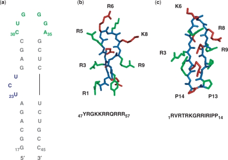
Figure 1.
(a) Sequence and secondary structure of HIV-1 TAR RNA; residues are colored showing the cyclin T1-binding site in green, the Tat-binding site in blue and the remaining double helical regions in gray. (b) Sequence of the 11-mer Tat-derived peptide corresponding to the Tat basic domain. (c) Cyclic peptide inhibitor of Tat–TAR complex. Both peptides are shown with side chains colored in alternating green and red with the peptide backbone colored in blue.

