Figure 3.
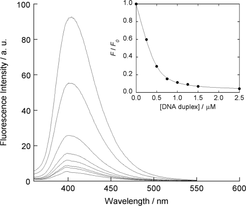
Fluorescence reponses of ATMND (500 nM) to 21-meric AP site-containing DNA duplex [5′-GCA GCT CCC GXG GTC TCC TCG-3′/3′-CGT CGA GGG CCC CAG AGG AGC-5′, X = AP site (dSpacer), C = target cytosine], measured in solutions buffered to pH 7.0 (10 mM sodium cacodylate) containing 100 mM NaCl and 1.0 mM EDTA. Excitation wavelength, 350 nm; temperature, 20°C. Inset: nonlinear regression analysis of the changes in the fluorescence intensity ratio at 403 nm based on a 1:1 binding isotherm model. F and F0 denote the fluorescence intensities of ATMND in the presence and absence of DNA duplexes, respectively.

