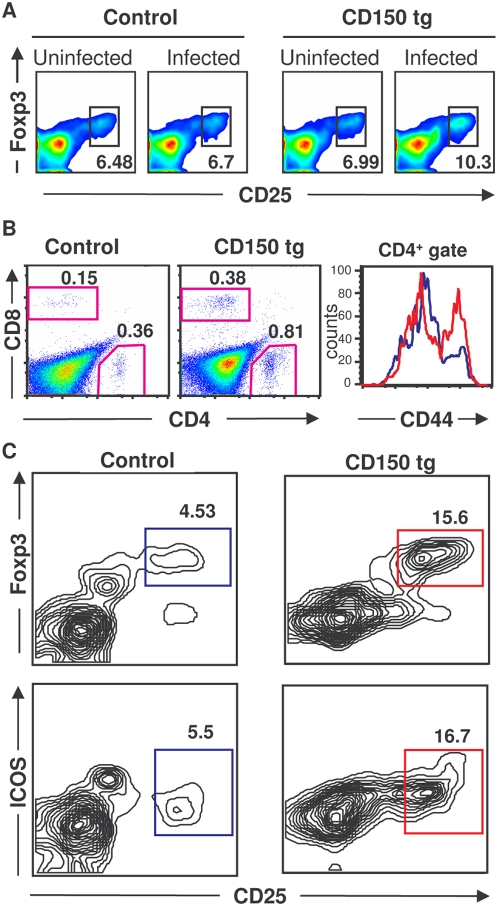
Figure 3. MV infection increases the proportion of CD4+CD25+Foxp3+ Tregs.
(A) Splenocytes from CD150 or nontransgenic mice (control), inoculated i.n. with either MV or medium (uninfected), were harvested 13 dpi and stained for CD4 and CD25 followed by anti-Foxp3 intracellular staining and analyzed by flow cytometry. (B, C) CD150×Foxp3-GFP transgenic mice and Foxp3-GFP littermates (control) were inoculated i.n. with MV. Brains were harvested 8 dpi and analyzed by flow cytometry as described in Methods. (B) Proportion of infiltrating CD4+ and CD8+ T lymphocytes in the brain (two left panels); expression of the CD44 activation marker on CD4+ T lymphocytes (right panel, CD150 transgenic in red and nontransgenic control in blue). (C) Tregs detected by the co-expression of Foxp3 and CD25 or ICOS. Results are representative of 4 independent experiments, each involving 4–7 mice per group. Differences between infected and noninfecetd mice were statistically significant (p<0.05, Student t-test).

