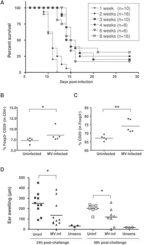
Figure 6. MV-induced immunosuppression in adult transgenic mice.
(A) Transgenic CD150×IFNα/βR KO mice were infected i.n. with MV at different ages and monitored for survival by Kaplan-Meier analysis. (B, C) Groups of 5 mice, 6 to 7 week-old, were infected i.n. with MV or left untreated. Splenocytes were harvested at 11 dpi and stained for CD4 and CD25 followed by anti-Foxp3 intracellular staining and analyzed by flow cytometry. Results are presented: (B) as the percentage of CD25+Foxp3+ cells within CD4+ compartment, for each analyzed animal and (C) as a percentage of CD25+ cells within the CD4+Foxp3+ population. Horizontal bars correspond to mean values. (D) Groups of 10 mice, 6 to 7 weeks old, were infected i.n. with MV or left untreated. Seven days later, mice were sensitized with DNFB 0.5% on the ventral skin or left unsensibilized (unsens). All mice were challenged 5 days later with DNFB 0.1% on the left ear. Results are presented as the individual ear swelling of two independent experiments and horizontal bars correspond to mean values. (** p<0.01, * p<0.05, Mann-Whitney test).

