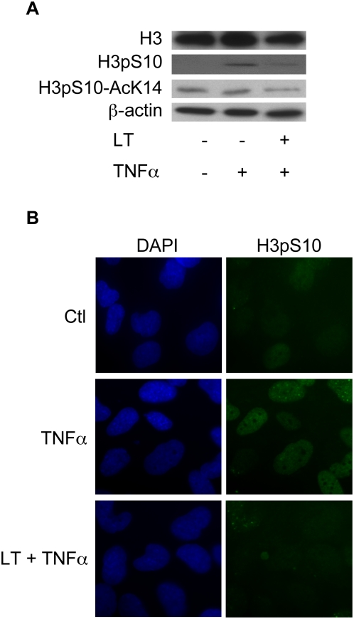
Figure 5. LT impairs H3 phosphorylation.
Beas-2B cells were incubated for 1 h with LT (1 µg/ml) and stimulated with TNFα (10 ng/ml) for an additional 2 h. Western blot analyses were performed using H3, H3pS10, and H3pS10-AcK14 antibodies and were normalized with a β-actin antibody (A). Immunofluorescence analyses were performed using an H3pS10 antibody and DAPI (B).

