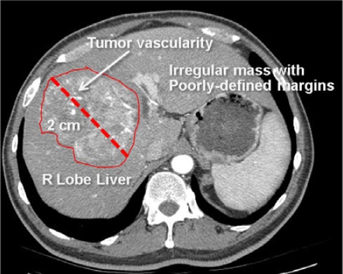
Figure 1.
Radiological image with semantic annotations drawn in an image annotation tool.
This image is annotated to convey the major semantic content, including anatomy (right lobe of liver), pathology (irregular mass, tumor vascularity), and imaging observations (2 cm in size, irregular shape, and poorly-defined margins). These image labels are human-readable, but not machine-accessible.

