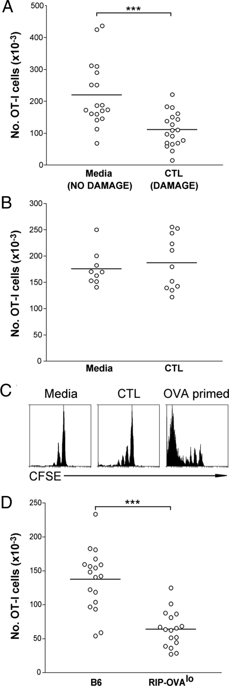
Fig. 4.
OVA released by CTL-mediated islet destruction induces deletion of naive OVA-specific cells. (A) RIP-OVAlo mice were given either media (NO DAMAGE) or 0.22 × 10⁶ Ly5.1+ CTLs (DAMAGE) i.v. followed by 5 × 10⁶ Ly5.2+ naive OT-I cells i.v. 4 days later. Four weeks after naive OT-I injection, the number of Ly5.1− OVA-tetramer+ cells (derived from the original naive Ly5.2+ OT-I cells) remaining in the spleen and lymph nodes of the mice was determined by flow cytometry. Pooled data are shown from 5 independent experiments. ***, P < 0.001. Circles represent individual mice and the bar represents the average. (B) An experiment was performed as in A except nontransgenic B6 mice were used as recipient mice instead of RIP-OVAlo mice. Pooled data are shown from 2 independent experiments. (C) A similar experiment to that performed in A was undertaken, with RIP-OVAlo mice given media alone (Left) or activated CTL (Middle), except that naive OT-I cells were labeled with CFSE and their proliferation was assessed after 4 weeks. As a positive control (Right), B6 mice were injected i.v. with 5 × 10⁶ naive CFSE-labeled OT-I cells followed by 2 × 10⁷ OVA-coated splenocytes (46) with 1 μg LPS (OVA primed) and analyzed after 4 weeks. Representative data from 11 mice per group collected over 2 independent experiments are shown. (D) B6 or RIP-OVAlo mice were given 0.22 × 10⁶ CTLs (i.v.). Four weeks after CTL injection, the number of CD8+OVA-tetramer+ cells remaining in the spleen and lymph nodes of the mice was determined by flow cytometry. Pooled data are shown from 3 independent experiments. ***, P < 0.001. Circles represent individual mice and the bar represents the average.

