Fig. 1.
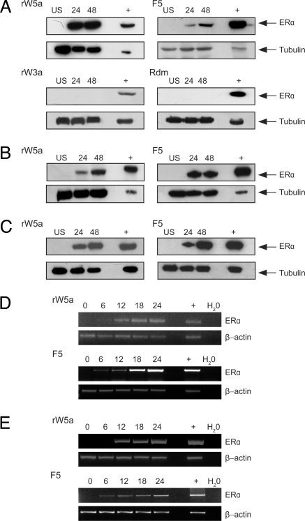
Wnt-5a signaling restores ERα expression. Breast cancer cells were grown in normal media and stimulated with rWnt-5a protein (rWnt-5a, 0.6 μg/ml), the Wnt-5a derived Foxy-5 peptide (F5, 100 μM), recombinant Wnt-3a protein (rW3a, 0.1 μg/ml), or a formylated random hexapeptide (Rdm, 100 μM) for 6–48 hours. After treatment, cells were lysed and subjected to SDS-PAGE, transferred to PVDF membranes, and blotted for ERα expression. (A) MDA-MB-231 cells stimulated with rWnt-5a, F5, rW3a, or Rdm. (B) MDA-MB-468 cells stimulated with rWnt-5a or F5. (C) 4T1 cells stimulated with rWnt-5a or F5. T47D cell lysates known to express ERα (+) were included to confirm the correct band size for ERα. Representative blots from at least three separate experiments are shown. (D) MDA-MB-231 cells stimulated with rWnt-5a or F5. RNA was extracted at the end time point, cDNA synthesized, and subjected to RT-PCR for ERα and the housekeeping gene β-actin. (E) RT-PCR for ERα and β-actin on RNA extracted from MDA-MB-468 cells stimulated with rWnt-5a or Foxy-5. In both (D) and (E), the positive control (+) is RNA extracted from T47D cells that express ERα, whereas the negative control is a water control. Representative agarose gels from three separate experiments are shown.

