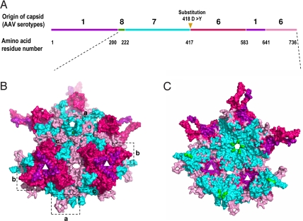
Fig. 1.
Sequence and structure analysis of the M41 capsid. (A) The primary structure of M41 capsid by alignments of its VP1 amino acid sequence with those of the parental AAV serotypes. A D to Y substitution on residue 418 is shown by a triangle. (B and C) The structural model of the 9-mer AAVM41 VP3 subunits reconstructed from the known crystal and homologous model structures of the parental viruses with the exterior surface (B), and interior surface (C), respectively. Sequences derived from AAV1 are colored in purple, AAV6 in hot pink and light pink (2 segments), AAV7 in cyan, AAV8 in green. The axes of symmetry are shown by white pentagon (5-fold), triangle (3-fold) and oval (2-fold) on the structural models. The structural regions around them are highlighted by frames a, b, and c.

