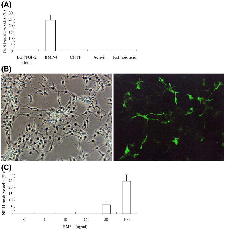
Figure 1.
Expression of NF-H by SFME cells. Cells were cultured for 4 d in DMEM/F12 medium with 0.5 ng/mL EGF/ FGF-2 alone, 100 ng/mL BMP-4, 100 ng/mL CNTF, 100 ng/mL activin, or 500 nM RA and then subjected to immunofluorescent staining for NF-H. (A) Percentage of cells staining positive for NF-H (mean±SD); (B) NF-H expression (green) in 100 ng/mL BMP-4 (left, phase contrast image; right, fluorescence image); (C) concentration dependence of BMP-4 on expression of NF-H.

