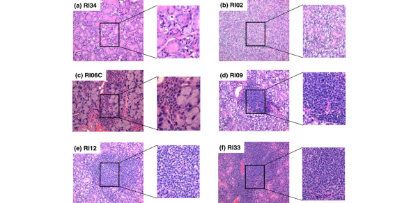
Figure 3.
Histological characterization of sialadenitis of male and female C57BL/6.NOD-Aec1Aec2R(n) mice. Submandibular glands were freshly explanted from male and female C57BL/6.NOD-Aec1Aec2R(n) mice euthanized at 20 or 24 weeks of age. The glands were fixed in 10% formalin, embedded in paraffin, and sectioned and stained with hematoxylin and eosin (H&E) dye. Representative H&E-stained histological sections of submandibular glands of selected recombinant inbred (RI) lines are presented: (a) RI34, (b) RI02, (c) RI06C, (d) RI09, (e) RI12, and (f) RI33. Original images were taken at × 100 magnification, with inserts expanded to show structural detail.

