
Figure 5.
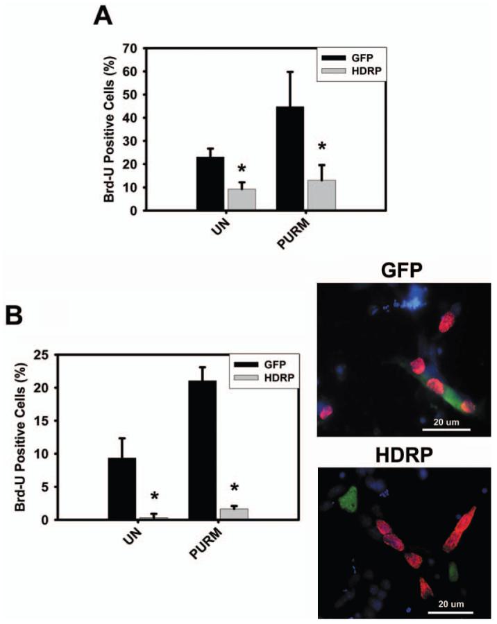
HDRP represses SHH-induced proliferation of HT22 and glial cells. (A) HT22 cells were grown in the presence of serum until 50% confluence when the cells were infected with HDRP or GFP containing adenovirus constructs. After 24 hours the medium was then changed to medium without serum and with or without (UN) 2 μM purmorphamine (Purm). Cells were incubated for an additional 48 hours, Brd-U was added to the medium for 2 hours, and immunocytochemistry performed. Brd-U and HDRP were detected using monoclonal antibodies against Brd-U and c-myc (HDRP contains a c-myc epitope tag) respectively. HDRP and GFP expressing cells were scored as Brd-U positive or negative and the results from three separate experiments were tabulated. (B) Glial cells were extracted from the cerebellum of rats and infected with HDRP or GFP containing adenovirus one day after plating. The following day the cells were treated with or without (UN) 2 μM purmorphamine (Purm). After 48 hours Brd-U was added for 2 hours and immunocytochemistry was performed. Cells expressing exogenous HDRP or GFP were scored as Brd-U positive or negative. The right panel shows a representative image of glial cells stained for GFP (green) or HDRP (green) and Brd-U (red). DAPI (blue) was used to visualize the nucleus and 20 μm scale bar shown for reference. * indicates statistical significance (P < 0.05) comparing GFP and HDRP groups for each treatment. A color version of this figure is available in the online journal.