
Figure 6.
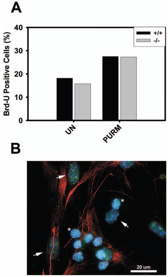
Proliferation of glial cells lacking HDAC9/HDRP. (A) Glial cells were extracted from HDAC9/HDRP null mice and wild-type littermates. The cells were treated with or without (UN) 2 μM purmorphamine (Purm) 48 hours after plating. Glial cells were then incubated for 48 hours, Brd-U was added to the medium for 2 hours, and immunocytochemistry subsequently performed. Brd-U was detected using monoclonal antibodies against Brd-U and glial cells were labeled using an antibody against GFAP. Glial cells were scored as Brd-U positive or negative and the results from two separate experiments were tabulated. (B) Endogenous HDRP (green) was detected in glial cells by immunocytochemistry using an antibody against HDRP. Glial cells (arrowheads) were positively identified using an antibody against GFAP (red). The smaller non-GFAP staining cells are CGNs (*). DNA was counterstained with DAPI (blue) to visualize the nucleus. A 20 μm scale bar is included for reference. A color version of this figure is available in the online journal.