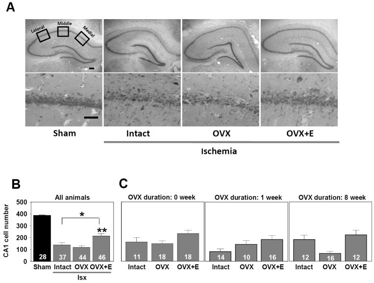
Figure 2.
Estradiol replacement modestly reduces CA1 cell loss in OVX, middle-aged females after global ischemia (Isx). (A) Representative photomicrographs from the 1 week OVX interval of intact, OVX, and OVX+estradiol (E) middle-aged female rats subjected to global ischemia or sham surgery. At 12 days after reperfusion, viable neurons in 4 toluidine blue stained sections containing the dorsal hippocampal CA1 were counted in 3 sectors (lateral, middle, and medial). Scale bars, Lower magnification (4X), 400 μm; higher magnification (40X), 60 μm. (B) Data represent a grand sum across 3 counting sectors and over right and left hemispheres. Because no statistical difference was found among the different sham groups (intact, OVX, OVX+E) at 1 and 8 weeks after OVX), these animals were pooled into one sham group. Global ischemia induced CA1 cell loss vs sham-operated animals. Treatment with E significantly increased CA1 cell survival. * p< .01 compared to sham-operated rats, ** p < .01 compared to intact and OVX rats. (C) There was no impact of OVX duration on CA1 cell survival.

