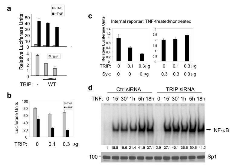
Figure 6. TRIP inhibits NF-κB activity.
(a) MCF7(BD) cells were transfected with NF-κB-luciferase reporter plasmids and increasing amounts (0.1 or 0.3 µg) of expression plasmids for wild-type TRIP (WT). Following treatment with or without TNF-α (20 ng/ml) for 18 h, relative luciferase units were measured. Basal NF-κB activities (−TNF) are expanded and shown in the lower panel. (b) MCF7(BD) cells were transiently transfected with a constitutively active luciferase reporter construct along with increasing amounts of the TRIP-expression vector. Cells were treated with or without TNF for 18 h and the amount of luciferase in the cell lysates was quantified. (c) Increasing amounts of the TRIP-expression plasmid were transfected into MCF7(BD) cells with 0.3 µg g of empty vector (left panel) or with 0.3 µg of the Syk-EGFP-expression vector (right panel). The luciferase activities of the constitutively expressed control reporter (pRL-TK) are shown. The Relative Luciferase Units represent the luciferase activities of TNF-treated samples divided by those of nontreated samples within the same group. (d) Nuclear extracts from control (Ctrl) and TRIP siRNA-transfected MCF7(BD) cells were prepared for the gel-shift assay. The shifted bands representing NF-κB complexes are indicated by the arrowhead. Their relative intensities were quantified and results are indicated below the panel. The nuclear extracts were analyzed by Western blotting with an anti-Sp1 antibody to show equal loading.

